Why focusing on large viruses?
Mimivirus genome surprises
"Bag of genes" vs reductive evolution
Understanding the origin of large DNA viruses
Why focusing on large viruses?
In summary, the prevalent view is that viruses were lucky combinations of self-replicating molecules - not living organisms - and per se not noble enough to deserve a genealogy.
The identification of many large DNA viruses exhibiting more than two hundred, then three hundred genes (Table 1) over the last 14 years, was apparently not sufficient to change this view, outside of a small circle of specialists.
With the discovery of mimivirus, the first virus that overlap with parasitic cellular organism in term of size (400 nm) [2] and genome complexity (over 1000 genes) [3], we now believe with have a unique opportunity to challenge the conservative attitude about viruses, and thoroughly revisit the concept and biological significance of virus (at least large DNA viruses). A useful first move would be to depart from the world of regular viruses by using a new name (for instance: Girus, Archevirus, ... etc) to refer to mimivirus and future other cell-sized viruses.
Mimivirus genome surprises
1)mimivirus is a "regular" nucleocytoplasmic large DNA virus (NCLDV): it contains all the core genes that have been identified as strictly or most often conserved in previously described pox-, irido-, asfar- and phycodna- viruses.
2)Yet, an analysis of the most similar homologs of mimivirus genes, as well as its pattern of loss of facultative NCLDV "core genes" does not suggest an affinity with one of the established NCLDV families. Mimivirus appears to be the first representative of the mimiviridae, a proposed new NCLDV group.
3)In addition to a normal complement of NCLDV gene homologs, mimivirus exhibits many genes encoding functions never encountered in any virus, including eight components central to protein translation: four aminoacyl tRNA synthetases (aaRS) together with four translation factors relevant to each of the initiation, elongation and termination steps. The enzymatic activity of mimivirus tyrRS has been experimentally verified.
This first encounter of translation apparatus protein encoding genes in a virus violates a well established dogma. Lacking ribosomes, viruses are supposed to entirely hand over the synthesis of their own proteins to their host's machinery. Already weaken by the discovery of numerous tRNA in some phycodnavirus [4], this dogma is now further demolished by the finding of virus-encoded translation enzymes and factors. Remember that aaRS are responsible for the correct application of the genetic code by insuring that the proper amino-acid is loaded on the right tRNA. The discovery of the first viral homologs of translation components also had important practical consequences. Like the DNA and RNA polymerases, aaRS have homologs within all domains of life: Archaea, Eubacteria and Eukarya. They could thus be used to improve the phylogenetic analysis of NCLDVs and precise their branching on the "Tree of Life", the root of which is the mythical Last Universal Common Ancestor (LUCA). The big surprise was then to see mimivirus defining its own branch, independent enough to suggest the existence of a 4th domain of life [Figure 1]. The precise scenario of what happened 3 billions years ago is a probably lost for ever, but the phylogenetic position of mimivirus (and by extension of other NCLDVs) is at least consistent with the hypothesis that the organism that provided mimivirus core gene set was already in existence at the time of (or prior) the emergence of the first eukaryotic cells. Such an ancestral anchoring of mimivirus to the Tree of Life is also consistent with previously proposed ideas linking DNA viruses to the emergence of the nucleus [5]. Thus, large DNA viruses could be promoted from the lowest status of non-living parasites to the one of bona fide ancestors of our own cells!
"Bag of genes" vs reductive evolution
Understanding the origin of large DNA viruses
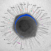
Figure 1. Mimivirus branching in the Tree of Life. None of the genes used to generate this tree exhibited evidence of recent lateral transfer.
| Organism | Code | Genome size (bp) | Date |
|---|---|---|---|
| Mimivirus | Y653733 | 1.181.404 | Nov 2004 |
| Treponema pallidum | NC_000919 | 1.138011 | Sep 2001 |
| Rickettsia prowazekii | NC_000963 | 1.111523 | Sep 2001 |
| Chlamydia muridarum | NC_002620 | 1.072950 | Oct 2001 |
| Chlamydia trachomatis | NC_000117 | 1.042.519 | Sep 2001 |
| Mycoplasma pulmonis | NC_002771 | 963.879 | Oct 22001 |
| Tropheryma whipplei | NC_004572 | 927.303 | Feb 2003 |
| Onion yellows phytoplasma | NC_005303 | 860.631 | Dec 2003 |
| Mycoplasma pneumoniae | NC_000912 | 816.394 | Apr 2001 |
| Mycoplasma mobile | NC_006908 | 777.079 | May 2004 |
| Ureaplasma parvum | NC_002162 | 751.719 | Jan 2000 |
| Wigglesworthia glossinidia | NC_004344 | 697.724 | Jul 2003 |
| Buchnera aphidicola | NC_004545 | 615.980 | Jan 2003 |
| Mycoplasma genitalium | NC_000908 | 580.074 | Jan 2001 |
| Nanoarchaeum equitans | NC_005213 | 490.885 | Feb 2004 |
| Canarypox virus | NC_005309 | 359.853 | Jan 2004 |
| Ectocarpus siliculosus virus | NC_002687 | 335.593 | Feb 2001 |
| Paramecium bursaria Chlorella virus 1 | NC_000852 | 330.743 | Feb 1996 |
| Shrimp white spot syndrome virus | NC_003225 | 305.107 | Nov 2001 |
| Human herpesvirus 5 | NC_001347 | 230.287 | Mar 1990 |
Table I. The largest viral genomes compared to the smallest procaryote genomes. Cellular organisms with a genome complexity lower than that of mimivirus are listed in red (redundant species are not listed). Polydnavirus is not listed, since its large (560 kb) atypical genome only encode 156 genes [6]. For all other organisms, there is approximately one gene per kb. A partial sequence (498 kb) of bacteriophage G genome (estimated length: 670 kb) is available from the Pittsburgh Bacteriophage Institute (http://pbi.bio.pitt.edu/).
REFERENCES
1.Chee MS, Bankier AT, Beck S, et al. Analysis of the protein-coding content of the sequence of human cytomegalovirus strain AD169. Curr. Top. Microbiol. Immunol. 1990 ; 154 : 125-169.2.Raoult D, Audic S, Robert C, et al. The 1.2-Megabase Genome Sequence of Mimivirus. Science Express 2004 (14 Oct 2004).
3.La Scola B, Audic S, Robert C, et al. A giant virus in Amoebae. Science 2003; 299: 233.
4.Van Etten JL, Graves MV, Müller DG, et al. Phycodnaviridae - large DNA algal viruses. Arch. Virol. 2002 ; 147: 1479-1516.
5.Pennisi E. The birth of the nucleus. Science 2004 ; 305: 766.
6.Espagne E, Dupuy C, Huguet E, et al. Genome sequence of a polydnavirus : insights into symbiotic virus evolution. Science 2004; 306: 286-9.